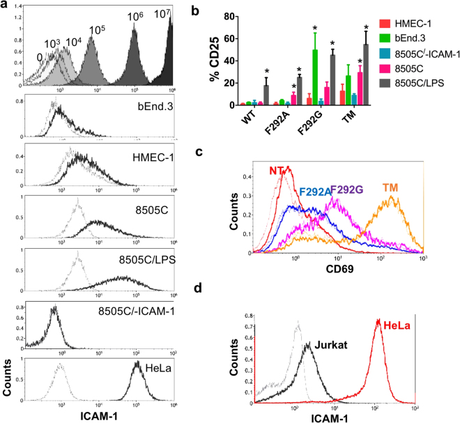
Figure 2.
Affinity and antigen-density dependent activation of Jurkat CAR T cells in vitro. (a) Top, histograms depicting 8 μm latex beads coupled with known amounts of R6.5 antibody conjugated with cy5.5 (103–107 antibodies per bead). The level of shift after incubation with R6.5 (black) from non-labeled (grey) was used to estimate ICAM-1 density in each indicated target cell line. 8505 C/-ICAM-1; 8505 C cells with ICAM-1 gene inactivation by CRISPR/Cas9. 8505 C/LPS, 8505 C cells were incubated with LPS to induce overexpression of ICAM-1. (b) CD25 expression in Jurkat CAR T cells (WT, F292A, F292G, and TM) after co-incubation with different target cell lines for 24 h (n = 3–4). p < 0.01 for * vs. 8505 C/-ICAM-1 by Dunnett’s multiple comparisons test. (c) Induction of CD69 after incubation with latex beads coated with 106 recombinant human ICAM-1-Fc molecules. Histograms correspond to CD69 expression in Jurkat cells without (thin, dotted line) and with (thick line) incubation with ICAM-1 coated beads. (d) ICAM-1 expression in Jurkat T cells compared to HeLa cells. Grey and black histograms correspond to unlabeled cells and R6.5 antibody-labeled cells, respectively.

