Figure 2.
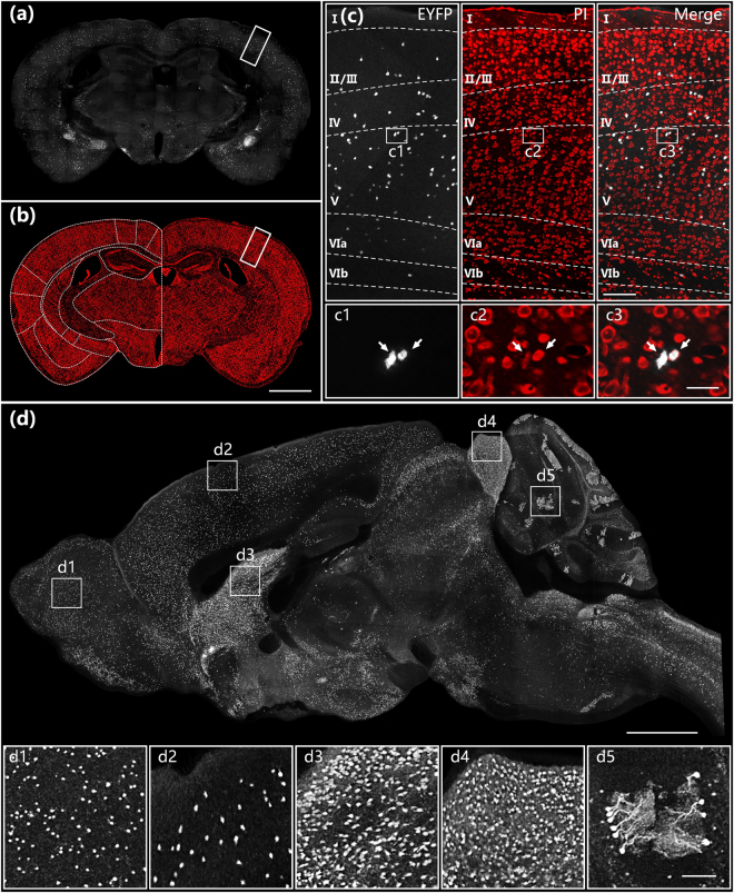
Whole-brain imaging of a SOM-IRES-Cre:Ai3-EYFP mouse brain. Images of EYFP-labelled neurons (a) and PI-stained cytoarchitecture (b) in the hippocampal coronal plane. (c) Enlarged views of white rectangular boxes indicated in (a) and (b) and their merged image. Image size is 900 × 400 μm. Projection thickness in the EYFP channel was 20 μm. (d) Sagittal reconstruction of maximum intensity projections of the SOM-IRES-Cre:Ai3-EYFP mouse brain. The projection thickness shown in (d) is 20 μm. The insets (d1)–(d5) are enlarged views of white square boxes shown in (d). The sizes of (d1)–(d5) are 400 × 400 × 20 μm. Scale bar: (a) and (b) 1 mm, (c) 25 μm, (c1)–(c3) 50 μm, (d) 1 mm and (d1)–(d5) 100 μm.

