Fig. 4.
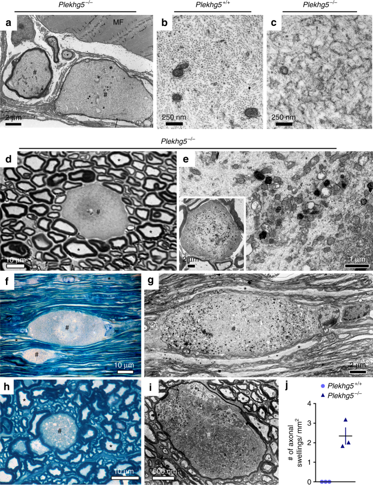
Loss of Plekhg5 impairs axonal integrity. a Swellings in distal motor axons. MF, muscle fiber. Scale bar: 2 µm. b, c High magnification micrographs of axons from wild-type b and Plekhg5-deficient c mice with altered cytoskeleton organization in Plekhg5 −/− mice. Scale bar: 250 nm. d Semi-thin cross-section of sciatic nerve from Plekhg5-deficient mice showing an axonal swelling. Scale bar: 10 µm. e High magnification micrograph of an axonal swelling from sciatic nerve. Scale bar: 1 µm. Inset, Scale bar: 2 µm. f–i Longitudinal- f, g and cross- h, i sections of lumbar spinal cord showing axonal swellings within the white matter of Plekhg5 −/− mice. f, h Scale bar: 10 µm. g, i Fine structure of axonal swellings in spinal cord white matter of Plekhg5-deficient mice. # labels axon swellings; asterisks label axons with unaltered morphology. g Scale bar: 2 µm. h Scale bar: 10 µm. i Scale bar: 500 nm. j Quantification of axonal swellings in spinal cord semi-thin cross-sections. Three animals per genotype were analyzed. Each data point represents the mean of 10 sections from individual animals with a distance of 100 µm between each section

