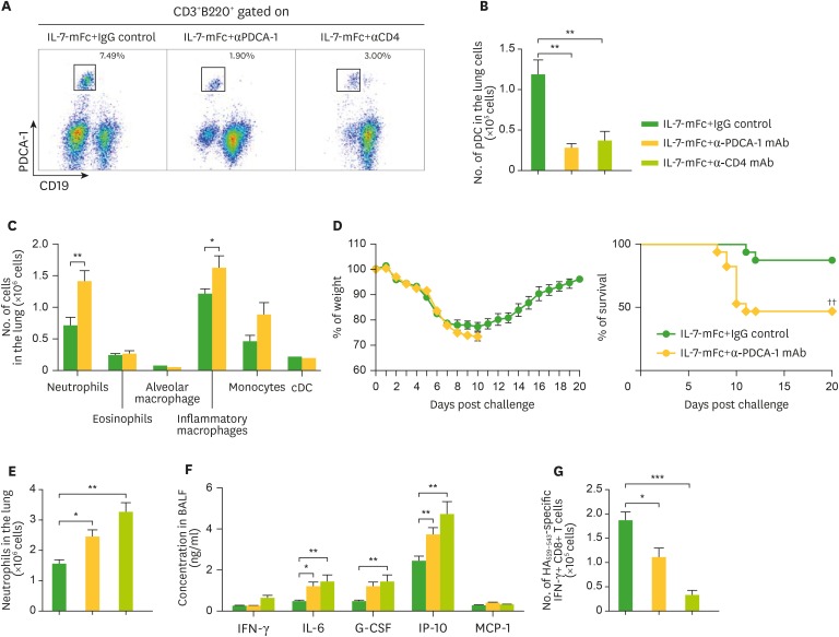
Figure 3.
Role of pDCs in IL-7-mFc-mediated protection against IAV. Mice (BALB/c, n=6) were treated with 200 μg of anti-PDCA-1, anti-CD4 antibody, or IgG control (D-1, D0, D4, D7) after IL-7-mFc treatment and challenge. (A-C) Representative plots and absolute number of pDCs and other myeloid cells in the lung of IL-7-mFc-treated mice measured 9 dpi. (D) Mice were challenged with a lethal dose of H5N2. Weight and survival of the mice were monitored daily. Absolute number of neutrophils (E) in the lung of IL-7-mFc treated mice and inflammatory cytokines and chemokines (F) at 9 dpi were also measured in the BALF. (G) Antigen-specific CD8+ T cell response was assessed by intracellular cytokine staining of IFNγ after HA529–543 stimulation. Results are representative of 2 independent experiment and expressed as the mean±standard error of mean.
*p<0.05, **p<0.01, ***p<0.001; ††p<0.01 by log-rank test.

