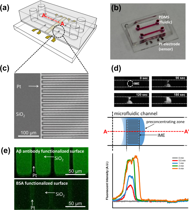
Figure 2.
The ICP preconcentrator-integrated sensor system. (a) The system was composed of 4 IMEs on a chip and 2 microfluidic channels for the ICP preconcentration phenomena. In a microfluidic channel, one IME was utilized for the antibody against the binding protein and the other IME was utilized for the reference electrode. (b) Image of the ICP preconcentrator-integrated sensor system with IME. (c) Scanning electron microscope image of IME. (d) Fluorescence images of Aβ preconcentration on IME, scheme of the ICP preconcentration phenomena on IME, fluorescent intensity of Aβ on IME and the location of the preconcentration plug over time. (e) Aβ antibody functionalization and BSA binding to block nonspecific binding.

