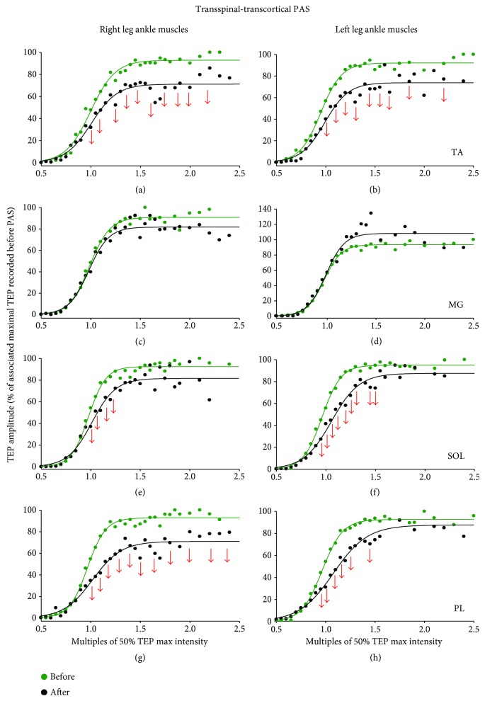
Figure 3.
TEPs recruitment curves before and after transspinal-transcortical PAS. Recruitment input-output curves of transspinal-evoked potentials (TEPs) recorded bilaterally from the TA, MG, SOL, and PL muscles from all subjects along with the sigmoid function fitted to the data. The abscissa shows multiples of stimulation intensities corresponding to 50% TEP max (S50). The ordinate shows TEP sizes as a percentage of the homonymous maximal TEP size obtained before transspinal-transcortical PAS. Red arrows indicate statistically significant differences (decreased amplitudes) before and after PAS based on repeated measures ANOVA. TA: tibialis anterior; MG: medial gastrocnemius; SOL: soleus; PL: peroneus longus.

