Figure 1.
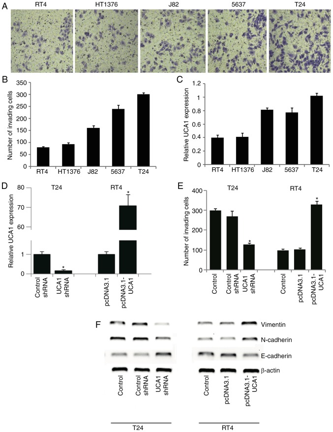
UCA1 modulated the invasion and EMT of bladder cancer cell lines. (A and B) Invasion capability of five bladder cancer cell lines was evaluated by Transwell assays. Crystal violet staining is shown in (A) and quantification of cells is shown in (B). (C) UCA1 mRNA expression levels in bladder cancer cell lines were evaluated by qRT-PCR. (D) T24 cells were transfected with UCA1 siRNA or control siRNA, and RT4 cells were transfected with UCA1-expressing pcDNA3.1 (pcDNA3.1-UCA1) or empty pcDNA3.1 vector (pcDNA3.1). Knockdown of UCA1 in T24 cells and overexpression of UCA1 in RT4 cells were validated by qRT-PCR. (E) Transwell assays in T24 and RT4 cells with UCA1 knockdown or overexpression. *P<0.05 vs. the control. (F) Western blot analysis of EMT markers E-cadherin, N-cadherin and Vimentin in response to UCA1 knockdown or overexpression. UCA1, urothelial carcinoma associated 1; EMT, epithelial-mesenchymal transition.

