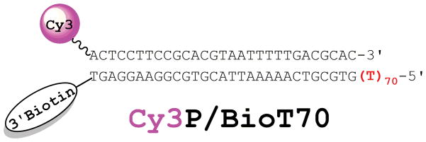
Figure 2.
Cy3P/BioT DNA substrate for SSB. The duplex region is identical to that described previously for RPA1 and the size (29 bp) is in agreement with the requirements for assembly of a PCNA ring onto DNA by RFC2,3. The length (70 nt) of the 5′ polyT ssDNA overhang shown in red will accommodate only a single SSB molecule at 200 mM ionic strength and permits RFC-catalyzed loading of PCNA. When pre-bound to Neutravidin, the biotin attached to the 3′-end of the template strand prevents loaded PCNA from sliding off the duplex end. A Cy3 dye attached to the 5′end of the primer strand serves as the FRET donor.

