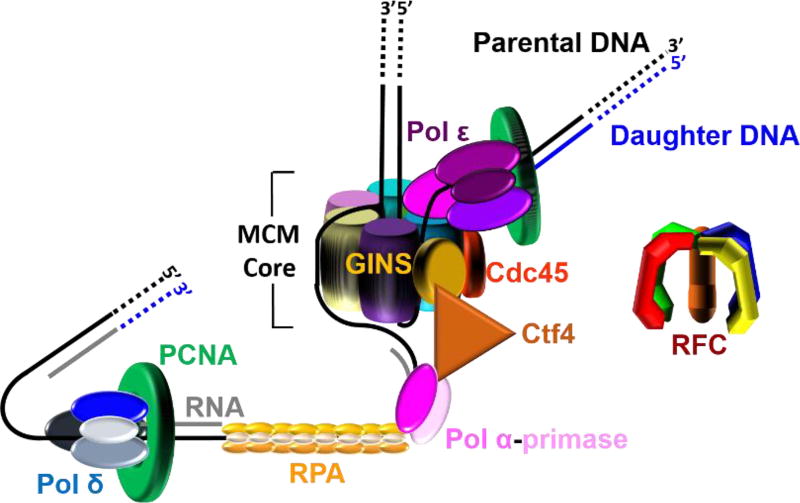
Figure 2.
A model of the eukaryotic replisome assembled at each replication fork. This model shown in cartoon form was generated from recent EM structures1, 2 and related studies (cited in main text). This model suggests that leading strand synthesis and at least the initiation of an Okazaki fragment occur on opposite sides of the helicase, necessitating an unexpected path for the templates as the dsDNA is unwound. The lagging strand template is sterically occluded from the central chamber of the MCM core and traverses the outside of the MCM core to reach pol α-primase. The leading strand template enters the CTD tier of the MCM core, traverses the central chamber or exits at an internal position, and then bends upward toward pol ε.

