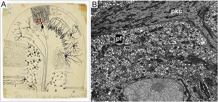
Fig. 4.

Two views of the cellular organization of the cerebellum. a Ramon y Cajal drawing of cell types of the cerebellum based on many observations of Golgi labeled neurons (Cajal Legacy, Instituteo Cajal, Madrid, Spain). Red box indicates relative scale of image in panel b. b Electron micrograph showing density of parallel fibers (pf; small, light circular profiles) surrounding the dendrites of a Purkinje cell (pkc; dark elongate profiles)
