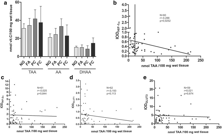
Fig. 5.

Expression of hypoxia marker HIF-1α associates with vitamin C level. (a) Analysis of ascorbic acid (AA), dehydroascorbic acid (DHAA) and both reduced and oxidized forms of ascorbate (TAA) levels in thyroid lesions (NG n = 51; FA n = 11; PC n = 23; FC n = 4). (b-e) Scatter plots showing HIF-1α (b), HIF-2α (c), hypoxia-related GLUT1 (d) and GLUT3 (e) proteins expression in relation to total vitamin C content in thyroid tumors (Spearman’s correlation). The median TAA value of 21,6 nmol/100 mg of tissue was used to classify sample as either ascorbate-deficient or ascorbate-replete. Ascorbate-deficient tumors had significantly higher levels of HIF-1 alpha protein than ascorbate-replete tumors (p < 0.01) by Mann-Whitney test
