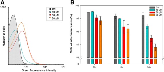
Fig. 3.

Determination of intracellular ROS accumulation and viability of cells exposed to 0–50 μM CuSO4. a Histogram showing ROS accumulation detected as green fluorescence from ROS-dependent oxidation of the ROS-sensitive probe H2DCF-DA following 30 min of incubation in the presence of Cu. The histogram shows data from a representative experiment. b Viability of cells following 2–24 h of exposure to CuSO4 measured as cells with intact membranes not stained by propidium iodide. Data are mean values from triplicate cultures from one representative experiment (the experiment was repeated twice). Error bars represents standard deviations
