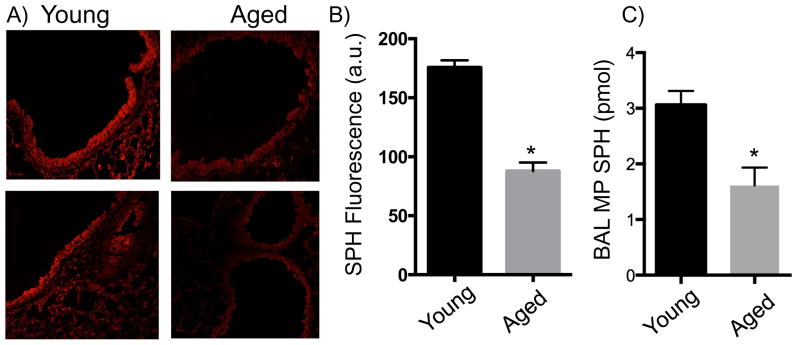
Figure 2. Decreased SPH in bronchial epithelium and BAL MPs in aged mice.
Fluorescent staining with anti-SPH antibodies of tracheal epithelial cells from A) young and B) aged mice. The top panels represent maximally observed SPH while bottom panels represent minimally observed SPH. C) Quantification of SPH in tracheal epithelial cells based on fluorescence. (n= 4–5 mice). D) Quantification of SPH in BAL MPs based on SPH kinase assay. (n=9–10 mice). Experiments were conducted twice to ensure reproducibility. *, p<0.05 as compared to young mice by Student’s t-test.

