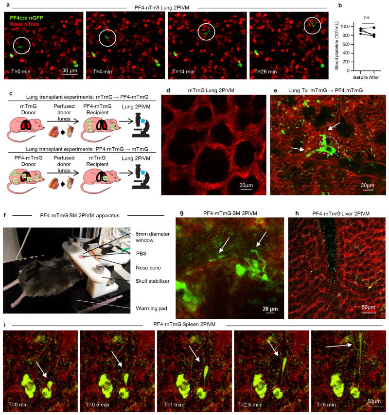
Extended Data Figure 1. MKs and proplatelets observed in lung circulation are from an extrapulmonary source.
(a) Lung 2PIVM of a PF4-nTnG mouse (nuclear GFP). The presence of the mobile GFP+ nucleated cells (circled) indicates the presence of a nucleus in circulating MKs. (b) Platelet counts in the blood before and after imaging. (c) Experimental schema of mTmG (perfused donor lung) to PF4-mTmG (recipient mouse) and vice-versa lung transplantation followed by 2PIVM. (d) 2PIVM of a mTmG mouse lung showing no GFP signal. (e) 2PIVM of a mTmG mouse lung transplanted into a PF4-mTmG recipient mouse showing GFP+ cells from recipient origin and platelet production in the lung. (f) BM 2PIVM apparatus. (g) Representative image of proplatelet release in the BM sinusoids (arrows). (h) Liver 2PIVM of PF4-mTmG mouse. Small platelets (GFP, green) were seen in the circulation but neither resident nor circulating MKs or proplatelets were observed. (i) Spleen 2PIVM of PF4-mTmG mouse. Sequential images show resident MKs (GFP, green) releasing proplatelets (arrows) in the spleen vasculature (in red).

