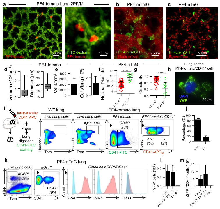
Figure 2. Resident MKs are present in the extravascular spaces of the lung.
(a-c) Visualization of resident/static MKs in the lung by 2PIVM of (a) PF4-tomato, (b) PF4-mTmG, or (c) PF4-nTnG mice. (d) Size characterization of PF4+ cells (red, >10μm) by quantitative image analysis of PF4-tomato lungs. Min-to-max boxplots are presented. (e) Quantification of PF4+ cells (red, >10μm). (f) Comparison of nuclear size and (g) circularity between PF4+ cells (green) and all other lung cells (red) by quantitative image analysis of PF4-nTnG lungs. Unpaired t-test: ****P < 0.0001. (h) Representative immunofluorescence images of PF4 and CD41+ cells sorted from perfused PF4-tomato lung and stained with anti-vWF (green) and DAPI (blue). (i,j) Intravascular (i.v.) or extravascular (e.v.) localization of PF4+ and CD41-FITC+ cells: (i) experimental schema, representative FACS plots, and (j) percentage (mean of 4 experiments, n=8 mice). (k) FACS gating strategy and surface expression of nucleated PF4+ and CD41+ cells from PF4-nTnG lungs. (l) FACS quantification of nucleated PF4+ and (m) nucleated PF4+/CD41+ cells present in PF4-nTnG whole lung, BM (2 femurs, 2 tibias × 6.6) and blood (1.5mL). (e-g,l,m) Mean ± S.D. are presented.

