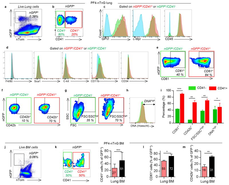
Extended Data Figure 3. Surface expression of lung MKs compared to BM MKs.
(a) Flow cytometric analysis of nGFP+ cells from PF4-nTnG lungs. (b) CD41 expression defines two populations of MKs: CD41+ (red) and CD41- (green). (c) Positive surface expression was detected for the following markers: GPVI, c-Mpl and CD45 in both populations. Unstained cells are plotted in blue. (d) Negative surface expression was detected for the following markers: F4/80, CD34, CD11b, Sca-1, c-Kit. (e) The CD41+ population has a higher percentage of CD61+ cells, (f) CD42b+ cells, (g) larger cells, and (h) higher DNA content, and summarized in (i). (j) Flow cytometric analysis of nGFP+ cells from PF4-nTnG BM. Compared to the lung, the BM nGFP+ population has a higher percentage of (k) CD41+ cells, (l) CD61+ cells, and (m) CD42b+ cells. Data are representative of three or more replicates. Mean ± SD are presented. Unpaired t-test: *P < 0.05, **P < 0.01, ***P < 0.001.

