Fig. 2.

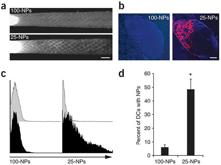
Ultra-small nanoparticles (25 nm) more efficiently drain to lymph nodes and are taken up by DCs than large nanoparticles (100 nm) upon intradermal injection. Fluorescently-labeled nanoparticles are seen draining through lymphatic capillaries (a; scale bar, 1 mm) and from isolated lymph nodes (b; scale bar, 200 μm; blue:cell nuclei, red:nanoparticles), with 25 nm particles trafficking more adeptly. Flow cytometry histograms (c) examined DC uptake of nanoparticles (black) or phosphate-buffered saline (grey) isolated from draining lymph nodes and was quantified (d). (For interpretation of the references to color in this figure legend, the reader is referred to the web version of this article.).
Reprinted by permission from Macmillan Publishers Ltd.: Nature Biotechnology [103], copyright 2014.
