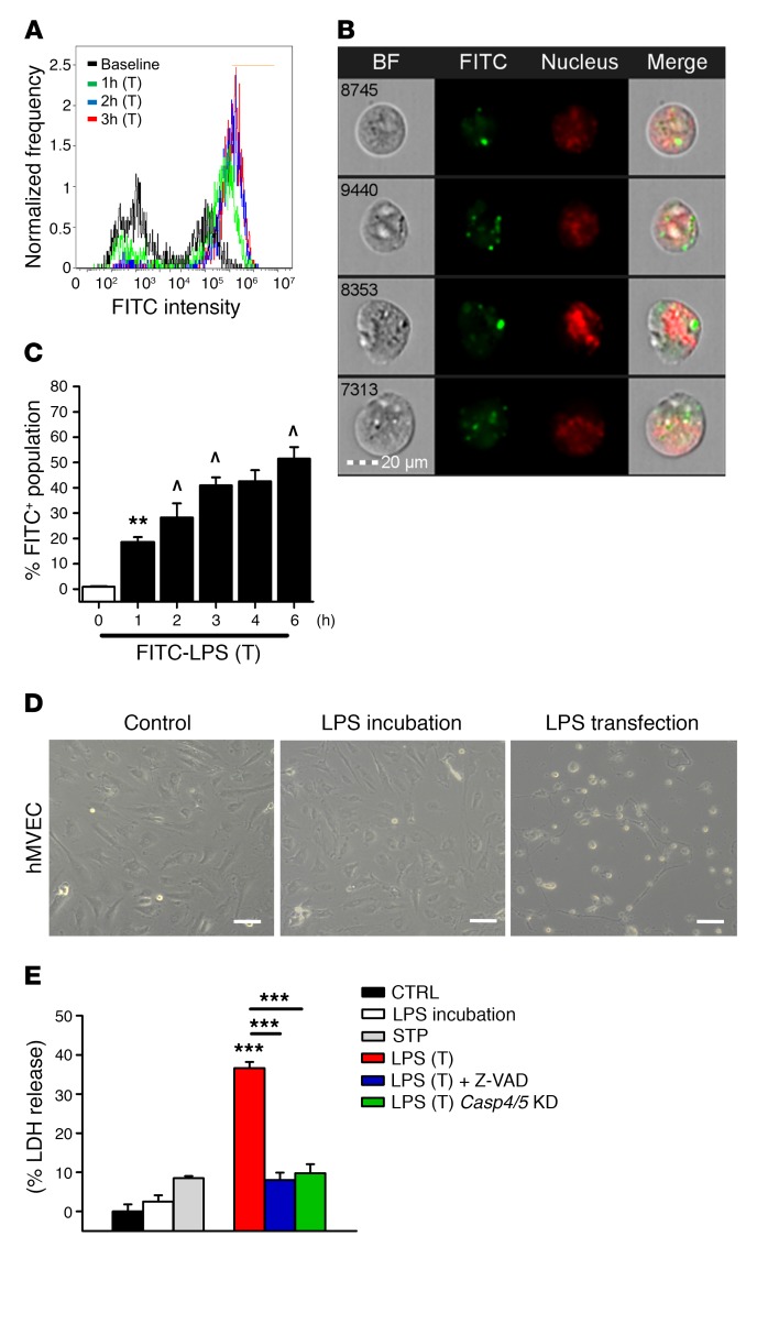
Figure 1. Intracellular LPS induces EC pyroptosis via activation of inflammatory caspases in mice and humans.
(A) Flow cytometry histograms and (B) representative cytometry images of hMVECs transfected with 2 μg/ml FITC-labeled LPS (O111:B4) for 3 hours. LPS fluorescence in green and nuclear staining in red show that LPS crossed the EC plasma membrane. Scale bar: 20 μm. BF, bright field. (C) Time course of hMVEC intracellular FITC-LPS fluorescence in the presence of a transfection (T) reagent. Results are shown as mean ± SEM. n = 5. **P < 0.01 from baseline; ^P < 0.01 from previous group using ANOVA. (D) Phase-contrast micrographs of control hMVECs after a 16-hour period of LPS (2 μg/ml) incubation and after a 16-hour period of LPS transfection (2 μg/ml) show that LPS transfection, but not LPS incubation, induced lytic cell death. Scale bars: 100 μm. (E) Release of LDH showed that LPS incubation (2 μg/ml) for 16 hours or staurosporine-induced (STP-induced) apoptosis resulted in minimal LDH release, whereas LPS transfection (2 μg/ml) led to marked cell lysis. Cells were primed with an initial exposure to LPS (500 ng/ml for 3 hours). Cell lysis was blocked by the pan-caspase inhibitor Z-VAD-FMK or knockdown (KD) of the human inflammatory caspases 4 and 5 (Casp4/5). Results are shown as mean ± SEM. n = 5. ***P < 0.001 using ANOVA.

