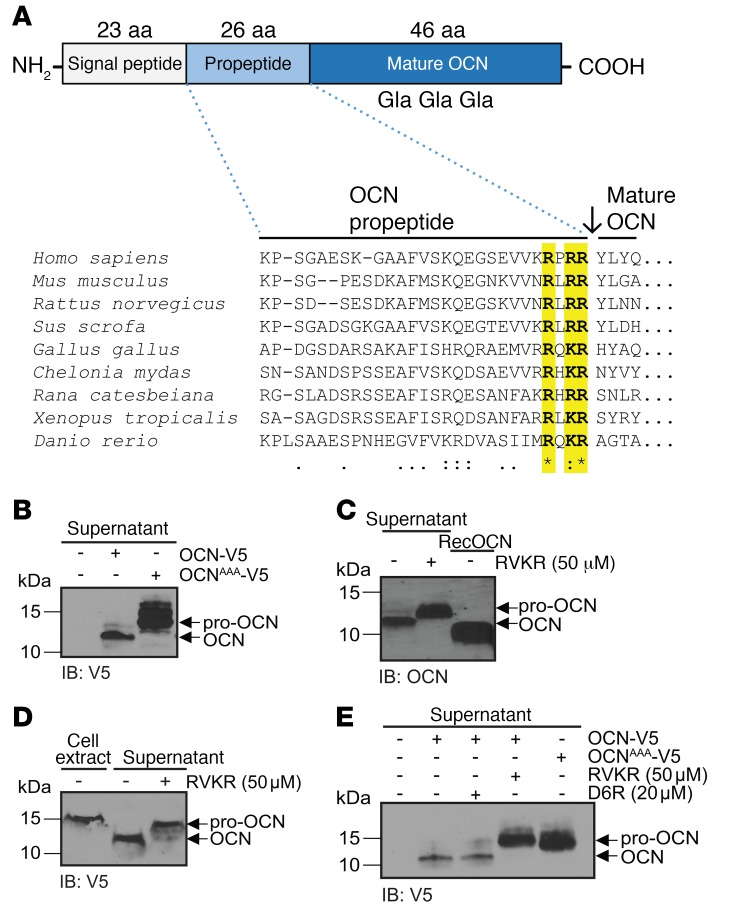
Figure 1. A PC cleaves pro-OCN at the RXRR motif in osteoblasts.
(A) Schematic representation of the pre-pro-OCN protein including the approximate positions of the Gla residues, and amino acid alignment of OCN propeptide sequences from various vertebrate species: the conserved RX(R/K)R motif is highlighted in yellow. Consensus symbols are included below the alignment. A single asterisk indicates a fully conserved residue; a colon indicates a strongly conserved residue; a period indicates moderate or weak conservation. (B) Western blot analysis of cell supernatant from primary osteoblasts transfected with OCN-V5 or an R46A/R48A/R49A OCN mutant (OCNAAA-V5), both tagged at the C-terminal with the V5 epitope. (C) Western blot analysis of endogenous OCN in the cell supernatant from differentiated mouse calvaria osteoblasts treated or not with 50 μM Dec-RVKR-CMK (RVKR). (D) Western blot analysis of cell supernatant and cell extracts of CHO-ldlD cells transfected with OCN-V5 and treated or not with 50 μM Dec-RVKR-CMK. (E) Western blot analysis of the supernatant of primary osteoblasts transfected with OCN-V5 or OCNAAA-V5 and treated or not with 50 μM Dec-RVKR-CMK or 20 μM D6R. IB, immunoblot.

