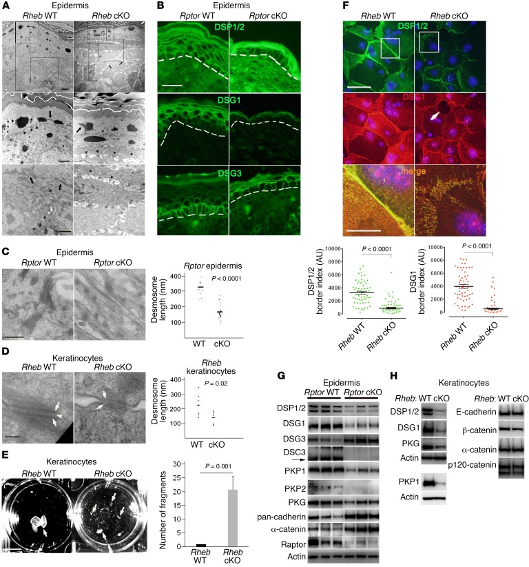
Figure 3. mTORC1 loss of function in the epidermis results in cell-cell adhesion defects.
(A) TEM of Rheb-cKO P0 epidermis (first row, E) demonstrates diminished granular layer (G) and intercellular gaps (first row, arrows). Desmosomes (second row, arrowheads) are evident (second row, arrow). Tonofilaments are collapsed (second and third rows, arrows) and not easily visible (third row, arrowheads) in cKO cells. Scale bars: 4 μm (top row), 2 μm (second and third rows). (B) Immunofluorescence for desmosomes (dotted line = dermal-epidermal junction). Representative images (r = 3). Scale bar: 30 μm. (C) Desmosomes in Rptor-cKO P0 epidermis have diminished keratin attachment by TEM (left panels; scale bar: 500 nm). Desmosomal length is quantified at right (bar represents mean length in nanometers; n = 12 and 29; P value by Student’s t test). (D) Desmosomes in Rheb-cKO keratinocytes have less dense cytoplasmic plaques (arrows) and poorly formed midline (arrowhead) (left panels; scale bar: 100 nm). Desmosome length is quantified at right (bar represents mean length in nanometers; n = 7 and 6; P value by Student’s t test). (E) Dispase dissociation for epithelial fragments (arrows) in Rheb WT and cKO keratinocyte monolayers (left panel). Scale bar: 1 cm. Quantification of dispase assay (right panel) (n = 4; error bars represent SEM; P value by Student’s t test). (F) Immunofluorescence for desmosomes in Rheb WT and cKO keratinocytes (top images), with intracellular gaps (arrow). Scale bar: 50 μm (top and middle images), 10 μm (bottom images). The third panel of merged staining is inset from the boxed areas in the top 2 panels. Quantification of desmosome protein border fluorescence intensity (bottom panels; r = 3, n = 58). Error bars represent SEM; P < 0.0001 by Mann-Whitney test. (G) Immunoblotting for junctional proteins in Rptor WT versus cKO P0 epidermis. (H) Immunoblotting for junctional proteins in Rheb WT versus cKO keratinocytes. PKP1 was immunoblotted separately using the same biological replicate.

