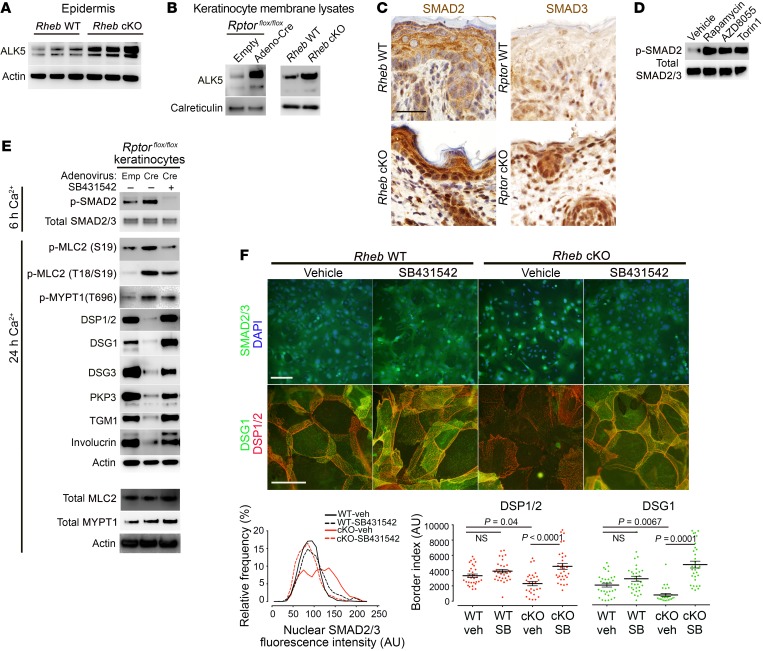
Figure 7. TGF-β receptor (ALK) expression and downstream signaling are upregulated in vivo and in vitro with mTORC1 loss of function and mediate increased ROCK activity.
(A) Immunoblotting of P0 Rheb WT and cKO epidermis for ALK5. (B) Immunoblotting of membrane lysates from Rheb WT and cKO keratinocytes and keratinocytes with or without inducible Rptor-cKO for ALK5. Calreticulin levels are used to normalize for membrane protein. ALK5 and calreticulin were immunoblotted in parallel, contemporaneously. (C) Immunohistochemistry for ALK activity markers in WT, Rheb-cKO, and inducible Rptor-KO P0 epidermis. Scale bar: 30 μm. (D) Immunoblotting of WT keratinocytes with or without mTORC1 (rapamycin, 200 nM) or mTORC1/2 inhibitors (AZD8055, 500 nM, or torin1, 1 μM) for markers of ALK activity. (E) Immunoblotting of lysates from WT or inducible Rptor-KO keratinocytes with or without ALK inhibitor treatment (SB431542, 10 μM) for markers of ALK activity, ROCK activity, and desmosome and differentiation markers. Total MLC2 and total MYPT1 were immunoblotted separately using the same biological replicate. (F) Immunofluorescence for SMAD2/3 localization (top row; scale bar: 100 μm) and desmosome proteins (middle row; scale bar: 50 μm) in Rheb WT and cKO keratinocytes with or without SB431542 (10 μM). Quantification of nuclear SMAD2/3 fluorescence (bottom row, left) from experiments above (r = 3, n > 1,874 cells, P < 0.0001 by Kruskal-Wallis test). Quantification of desmosome protein border fluorescence (bottom row, right) (r = 3, n > 28 cells, P values indicated are by 1-way ANOVA, DSP, or Kruskal-Wallis test, DSG1).

