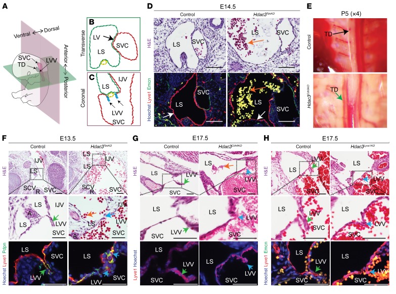
Figure 2. Hdac3 is an important regulator of lymphovenous valve development.
(A–C) Schematic model depicting normal anatomy of a developing murine lymphovenous valve (A, black arrow) in transverse (B) and coronal (C) planes. (D) Transverse sections of E14.5 Hdac3TekKO embryos revealed blood-filled lymph sacs (orange arrows) lined by lymphatic (Lyve1 immunostaining [red], white arrows), but not venous (Emcn immunostaining, green), endothelial cells compared with that seen in controls. (E) Dissected P5 Hdac3Cdh5KO mice had a blood-filled thoracic duct (green arrow) compared with a chyle-filled thoracic duct in control mice (black arrow). (F) H&E-stained coronal sections of an E13.5 Hdac3TekKO embryo revealed a blood-filled lymph sac (orange arrow) and disrupted morphology of the lymphovenous valves (green arrows) compared with controls (yellow arrows). Immunofluorescence staining for podoplanin (Pdpn) (green) and Lyve1 (red) showed overlapping expression (yellow) in E13.5 LVVs. Orange arrow indicates a blood-filled lymph sac. (G and H) H&E-stained coronal sections of E17.5 Hdac3Cdh5KO (G) and Hdac3Lyve1KO (H) embryos revealed disrupted morphology of the lymphovenous valves (blue arrows) compared with controls (green arrows). Orange arrow shows a blood-filled lymph sac. Lyve1 (red) was expressed in E17.5 murine lymphovenous valves (G and H). Emcn (green, venous marker) was used as a negative control for lymphovenous valves (H). IJV, internal jugular vein; LS, lymph sac; LVV, lymphovenous valve; SVC, superior vena cava; TD, thoracic duct. Scale bars: 100 μm and 50 μm (F, bottom panels, G, and H). See also Supplemental Figure 5 and Supplemental Table 5.

