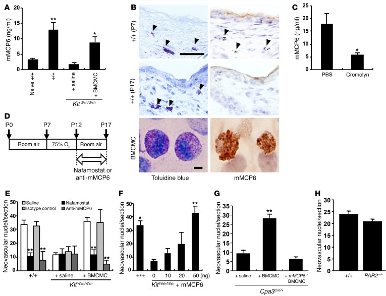
Figure 5. mMCP6 triggered retinal neovascularization.
(A) mMCP6 ELISA was performed using serum from the OIR model and age-matched naive WT pups on P17. n = 8 in each group. *P < 0.05; **P < 0.01 versus saline-injected mice, Dunnett’s test. (B) mMCP6 immunohistochemical analysis was performed on skin sections of WT mice on P7 and P17 and on BMCMCs cultured for 5 weeks. Results are representative of 3 independent experiments. Arrowheads indicate mast cells in the skin. On P17, mast cells were degranulated and mMCP6 positivity was not identified. (C) mMCP6 ELISA was performed using mouse serum obtained at 6 hours after treatment with cromolyn on P12. n = 8 in each group. *P < 0.05 versus control mice, Mann-Whitney U test. (D) Schematic of the study design. Pups were i.p. injected with 20 μl NM or anti-mMCP6 mAbs every day from P12 to P17. Animals were sacrificed, and eyes were enucleated on P17. (E) Quantification of neovascular nuclei in mice treated with NM and anti-mMCP6 mAbs. n = 9 in each group. **P < 0.01 versus saline-injected mice, Dunnett’s test. (F) Quantification of neovascular nuclei in mice injected with recombinant mMCP6. n = 9 in each group. *P < 0.05; **P < 0.01 versus KitWsh/Wsh mice treated with vehicle alone, Dunnett’s test. (G) OIR was not induced by reconstitution of Cpa3Cre/+ mice with mMCP6-deficient BMCMCs. n = 8 in each group. **P < 0.01 versus saline treatment, Dunnett’s test. (H) Retinal neovascularization in relative hypoxic condition in PAR2-deficient mice. n = 8 in each group. Scale bars: 50 μm (B, skins); 20 μm (B, BMCMC). Results are shown as mean ± SEM of values determined from 3 to 4 independent experiments (A, C, E–H).

