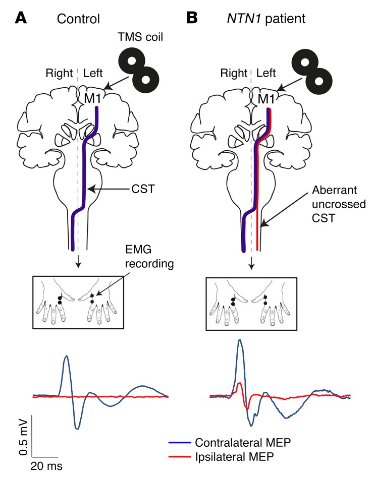
Figure 4. Analysis of neural signal propagation along the CSTs using single-pulse TMS.
(A and B) Schematic representation of the TMS experiments. In controls (A), unilateral stimulation of the hand area of the dominant primary motor cortex (M1) with TMS elicited contralateral MEPs only (A, blue line), whereas bilateral MEPs were observed in NTN1 patients (B, contralateral blue and ipsilateral red lines).

