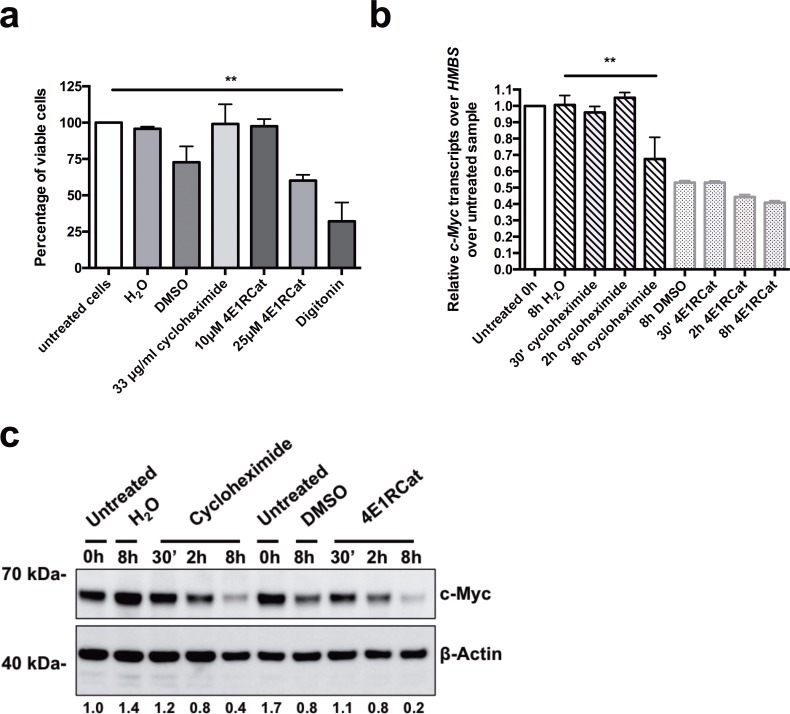
Fig 2. Treatment of BL cells with the mRNA translation inhibitors cycloheximide and 4E1RCat inhibits c-myc protein, but not c-myc mRNA expression.
(a) Akata cells were treated with the protein synthesis inhibitors cycloheximide (33 μg/ml) or 4E1RCat (10 μM and 25 μM) for 8.5h. Cell viability was determined by Trypan blue exclusion assay. Untreated and vehicle-treated cells (H2O for cycloheximide and DMSO for 4E1RCat) were used as negative controls, and digitonin (30 μg/ml) treated cells as a positive control for cell toxicity. Results are shown as percentage of viable cells (n = 3). (b) and (c) Akata cells were treated for 8h with the protein synthesis inhibitors cycloheximide (33 μg/ml) or 4E1RCat (10 μM) for 30 min, 2h or 8h or with their vehicles, H2O or DMSO, respectively. (b) The BZLF1 mRNA level relative to HMBS was measured by RT-qPCR and normalized to untreated samples. One representative experiment out of three is shown. (c) C-Myc protein expression level was measured by Western Blot. Densitometry of the c-myc protein expression was normalized over β-Actin expression and to untreated samples. Densitometry data of untreated cells was set to 1. Data are represented as mean ± SD. Statistics were calculated using the Tukey’s multiple comparison test (ANOVA). (*, P<0.05).

