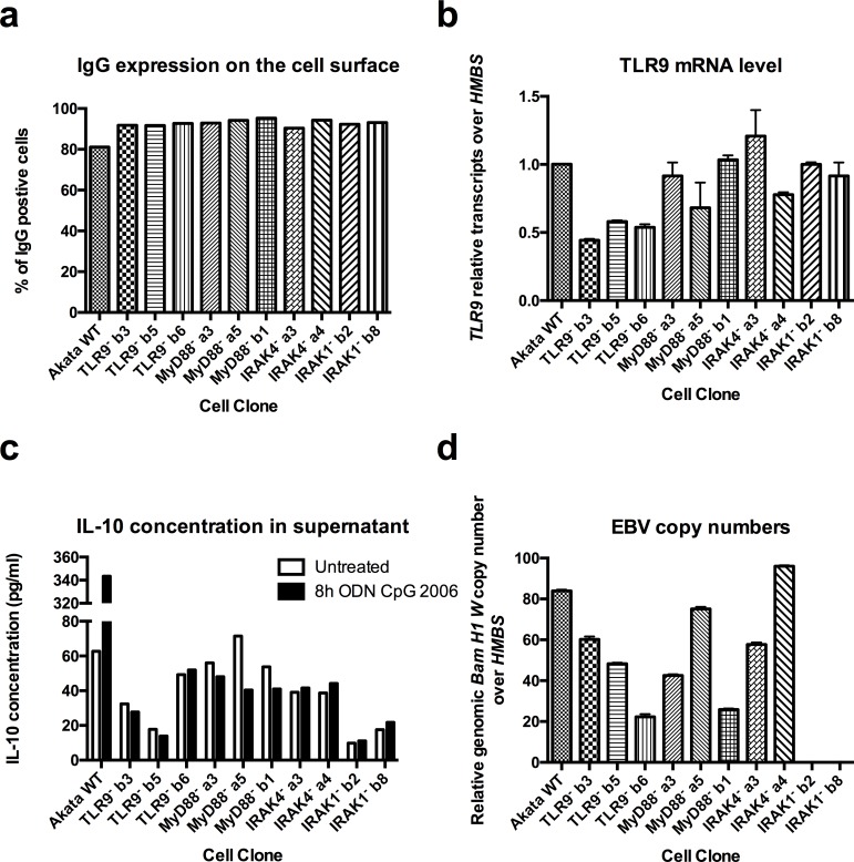
Fig 4. CRISPR/Cas9 inactivation of TLR9, MyD88, IRAK4 and IRAK1 results in a complete abrogation of TLR9 signaling.
Akata Burkitt’s lymphoma cells transfected with px458 plasmids coding for Cas9 and for sgRNA targeting TLR9 (TLR9-b3, TLR9-b5 and TLR9-b6), MyD88 (MyD88-a3, MyD88-a5 and MyD88-b1), IRAK4 (IRAK4-a3 and IRAK4-a4) and IRAK1 (IRAK1-b2 and IRAK1-b8), respectively, were diluted for single cell cloning, sequenced and clones containing an early stop codon were selected for further characterization. (a) Percentage of IgG positive cells was measured by flow cytometry. (b) TLR9 mRNA level was measured by RT-qPCR and normalized over HMBS and over WT Akata cells. WT Akata cells were set to 1. (c) IL-10 cytokine expression level measured by ELISA in the supernatant of untreated cells, and cells treated for 8h with ODN CpG 2006. (d) Viral BamH1 W copy numbers over cellular HMBS were determined by qPCR. (a) Has been performed once. (b, c, d) Shown is one representative experiment out of three. Data are represented as mean ± SD.

