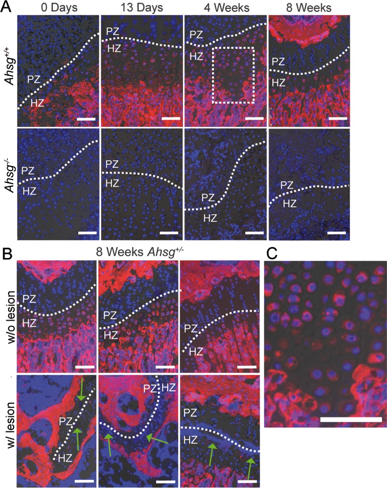
Fig 4. Growth plate histology and immunofluorescent localization of fetuin-A in distal femoral growth plates.
(A) Decalcified femur paraffin sections from mice of different ages were stained with an anti-fetuin-A antibody (red) and nuclei were counterstained with DAPI (blue). In the growth plate, fetuin-A was localized in hypertrophic chondrocytes. Ahsg-/- mice served as negative control (lower panel). (B) Similar to wildtype mice, fetuin-A was detected in hypertrophic chondrocytes from eight-week-old Ahsg+/- mice. Fetuin-A staining was negative in growth plates containing a lesion (green arrows). (C) A magnified view of the marked area in (A) shows the cytoplasmic localization of fetuin-A in hypertrophic chondrocytes. Scale bars are 75 μm.

