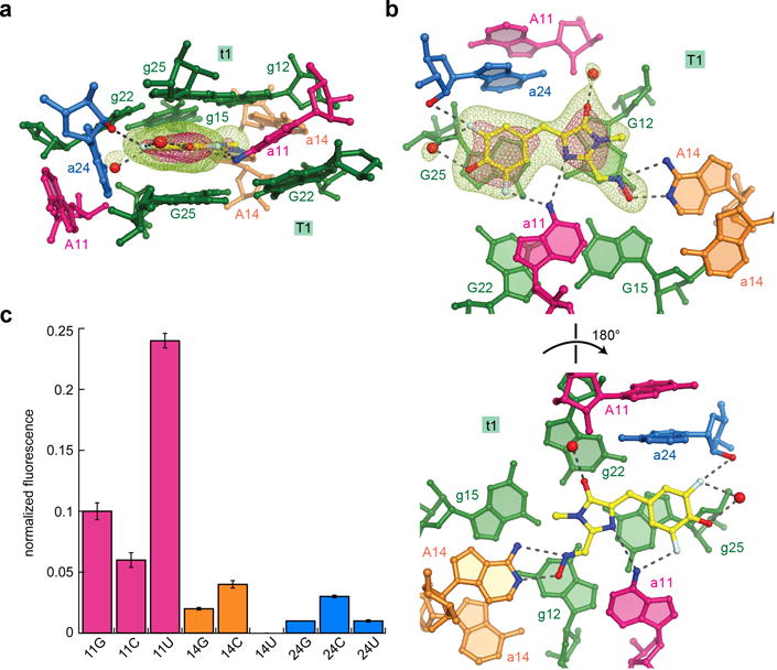
Figure 4.

Architecture and functional importance of quasisymmetric DFHO binding site. (a) Side view of the DFHO binding site, color-coded as in Fig. 1. Water molecules are depicted as red spheres. Mesh depicts a portion of the |Fo| − |Fc| electron density map, calculated before addition of DFHO and associated water molecules to the crystallographic model; green and pink mesh are contoured at 3σ and 7σ, respectively. (b) Axial views of the DFHO binding site, depicting DFHO above the T1 G-quartet, looking down on the T1 and t1 G-quartets (top and bottom, respectively) (c) DFHO fluorescence activation by point mutants of the three interfacial adenosines of Corn. Fluorescence of each mutant is normalized to that of the wild-type. Error bars represent s.d. from three independent experiments.
