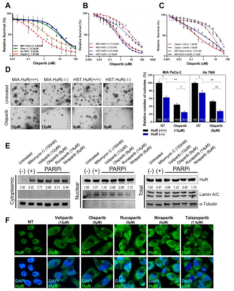
Figure 1. HuR expression regulates sensitivity to PARPi in PDA cells.
Cell survival of PDA cell lines (A), HuR-knockout CRIPSR cell lines, MIA PaCa-2 and Hs 766T [HuR(+/+) vs HuR(−/−)] (B) and HuR-silenced MiaPaCa-2 and Capan-1 cells (C) treated with increasing doses of olaparib for 7 days. (D) Representative images of MIA.HuR(+/+) vs MIA.HuR(−/−) and HST.HuR(+/+) vs HST.HuR(−/−) cells seeded and cultured in soft agar in the presence of respective IC50 doses of olaparib for 4 weeks. (E) HuR expression in MIA PaCa-2 cells treated with indicated IC50 doses of PARPi for 12hr, and fractionated as indicated. Lamin A/C and α-Tubulin used as controls to determine the integrity of nuclear and cytosolic lysates respectively. Mitomycin C used as positive control for cytoplasmic translocation of HuR. (F) Immunofluorescent images of HuR (green) in MIA PaCa-2 cells treated with PARPi for 12hr. Nuclei were stained with DAPI. Magnification 40X.

