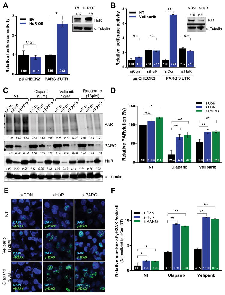
Figure 3. HuR regulates PARG protein expression and function.
Luciferase activity in MIA PaCa-2 cells co-expressing a luciferase reporter construct with PARG 3′UTR and (A) HuR overexpression or (B) HuR silencing (C) HuR, PARG and PAR protein expression in total lysates from HuR- and PARG-silenced MIA PaCa-2 cells treated with IC50 doses of indicated PARPi for 24hours, using α-Tubulin as a loading control. (D) ELISA indicating relative PARylation in MIA PaCa-2 cells transfected and treated as above. The indicated fold changes are means of three independent experiments, normalized to control transfected sample under no treatment (NT). (E) DSBs assessed by immunofluorescence staining for γH2AX (green) in MIA PaCa-2 cells transfected and treated as described above. (F) DNA damage foci were quantified and plotted ± SD.

