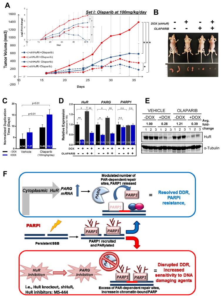
Figure 6. HuR silencing in vivo enhances olaparib- mediated suppression of PDA xenograft growth.
Mia.shHuR xenografts in athymic, nude mice were randomized into DOX and olaparib treatment groups. (A) Tumor volumes are plotted, with each point representing the mean ± 2SE of each group, *P<0.05. Inset shows differences in number of duplications. (B) Representative image of mice and tumor per group. (C) Tumor duplication time (days) per group (D) HuR, PARG and PARP-1 mRNA expression in extracted tumors, relative to vehicle- treated –DOX group. Each bar represents the mean ± SEM (n = 3 per group). (E) HuR protein expression when tumors were harvested (day 36, n=3). (F) Working model: In response to PARPi stress, cytoplasmic HuR binds to and stabilizes PARG mRNA, thereby increasing PARG expression and modulating PARP1-chromatin dynamics. HuR and PARG inhibition breaks such acute resistance by enhancing chromatin- trapped PARP-1 and accumulation of damaged DNA and apoptosis.

