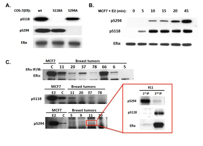
Figure 1. Specific immunoreactivity of pS294, its induction kinetics relative to pS118 in an ER-positive breast cancer cell line (MCF7), and the variable expression of endogenous pS294 found in ER-positive human breast tumors.

A. To demonstrate the immunospecificity of a newly developed anti-ERpS294 rabbit monoclonal, COS-7 cells were transfected with either wildtype (wt) ERα, ERα mutated at S294 (S294A) or ERα mutated at S118 (S118A); 24 h after transfection, media was changed to charcoal-stripped serum containing media for 24 hours, followed by E2 treatment at 10 nM for 45 min, and cells were then lysed, ERα immunoprecipitated and probed by western blotting. B. To compare the ligand induction kinetics of pS294 relative to pS118, MCF7 cells grown for >24 h in charcoal-stripped media were treated with E2 (10 nM) for the indicated times, ERα was immunoprecipitated and probed via western blot for pS294, pS118, and total ERα. C. To compare detection of pS294 expression in representative ER-positive primary breast tumor samples (#5, 6, 9, 11, 20, 37, 66, 78) relative to control (C) or E2 (45 min) treated MCF7 cells, whole cell lysates were first immunprecipated (IP) for total ERα. and further immunoblotted (IB) for pS294, pS118, and total ERα. As shown in the inset for tumor sample #11, parallel lysate aliquots were first immunoprecipitated for pS294 and that 1st IP immunblotted for pS294, pS118, and total ERα content (1st IP), while the remaining unprecipitated lysate was then immnoprecipitated for total ERα and immunoblotted for pS294, pS118, and total ERα (2nd IP).
