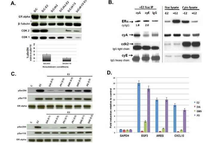
Figure 2. ERα ligand binding triggers rapid association with cyclin A/E-associated CDK2, whose suppression or enzymatic inhibition not only prevents pS294 formation but also the transcription of ERα inducible genes (EGF3, AREG, CXCL12).

A. Knockdown of CDK1 and/or CDK2 was performed on replicate wells of MCF7 cells transfected with either control (C), CDK2 or CDK1/2 targeted siRNA; 24 h later cultures were changed to phenol red-free media containing 10% charcoal-stripped serum and allowed to grow for another 24 h before treatment with E2 (10 nM x 30 min), followed by cell harvesting, protein extraction and western blotting for ERα, β-tubulin, CDK1 and CDK2 as shown. In parallel, immunoprecipitation of total ERα from the cell lysates was followed by western blotting to detect pS294 and pS118 levels; and densitometry measured band intensities were used to quantitate pS294 levels relative to pS118 levels after knockdown of CDK2 alone (SiCdk2) or combined CDK1/2 knockdown (SiCdk1/2) under E2 exposure, relative to control siRNA (SiC-E2) treatment conditions. The average relative declines (n = 3, SEM) in pS294 (relative to pS118) are shown in the bar graphs below. B. MCF7 cells grown in charcoal stripped and phenol red free media were treated +/- ligand (-E2 or +E2, 10 nM x 30 min), gently lysed and nuclei pelleted and extracted, producing cell fractions (Nuc lysate, Cyto lysate) that were immunoblotted for ERα, cyclin A2 (cyA), cyclin E (cyE) or CKD2 as shown. In parallel, the E2 treated Nuc lysates were first immunoprecipitated using anti-cyA, anti-cyE, or control anti-IgG and then immunoblotted for ERα, cyA, cyE and CKD2. C. MCF7 cultures pretreated for 60 min with either vehicle (C) or the indicated dose (μM) of CDK inhibitor (DIA = Dinaciclib, JNJ = JNJ7706621, BMS = BMS265246, SNS = SNS-032, PD = PD0332991/Palbociclib) were then stimulated for 20 min with E2 before harvest, protein extraction, ERα immunoprecipitation, and immunoblotting for pS294, pS118, and total ERα as shown. D. MCF7 cells grown in charcoal stripped and phenol red-free media were pretreated for 1 h with 0.5 μM DIA , 1 μM PD, or 1 μM BMS followed by 6 h E2 (10 nM) treatment, after which total RNA was extracted and semiquantitative RT-PCR performed to measure fold induction of the ERα inducible genes EGF3, AREG, and CXCL12 relative to the housekeeping gene GAPDH, using previously described primers and methods [11].
