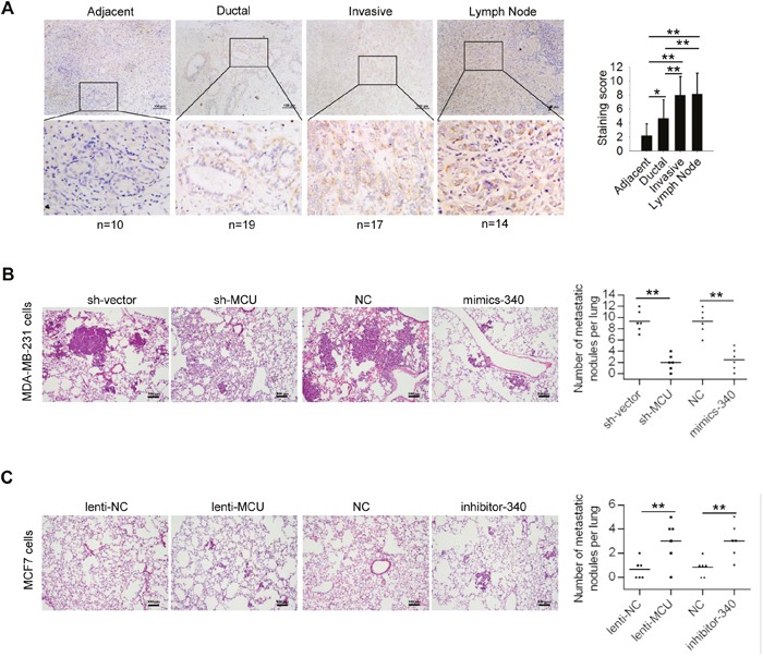
Figure 6. MCU and miR-340 expression regulate breast cancer metastasis in vivo.

(A) (Left) Immunohistochemical stains of representative samples of human breast carcinoma sections showing MCU expression. Scale bars (top row): 100 μm; inset (bottom row) scale: 50 μm. (Right) Immunohistochemical staining scores for the tissue samples examined: tissue adjacent to the tumor (n=5), ductal cancer tissue (n=14), invasive cancer tissue (n=12), and lymph node tissue (n=9). (B) Hematoxylin and eosin staining of representative lung tissue sections from mice injected with MDA-MB-231 harboring sh-MCU, miR-340, or NC. (C) Hematoxylin and eosin staining of representative lung tissue sections from mice injected with MCF7 breast cancer cells harboring lenti-MCU, inhibitor-340, or NC. Scale bars: 100μm. The number of lung metastatic foci in each group was determined in five randomly selected fields. The error bars in all the bar graphs represent SD. Statistical significance was determined via one-way analysis of variance followed by pairwise t-tests.*P< 0.05; **P< 0.01.
