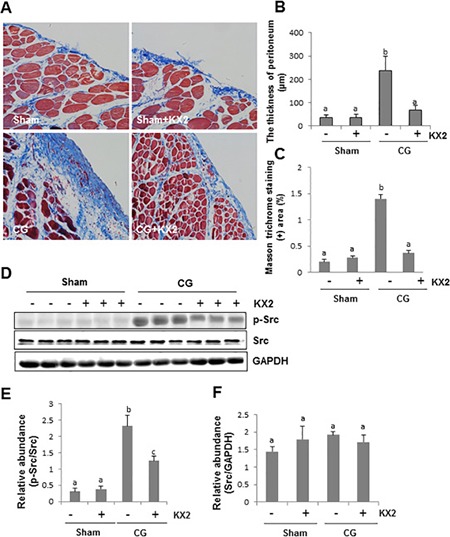
Figure 2. KX2-391 attenuates development of CG-induced peritoneal fibrosis.

Peritoneal membrane was collected at 21 days after CG injury with or without administration of KX2-391 (KX2) (A–F). (A) Photomicrographs illustrate Masson trichrome staining of the peritoneum. (B) The graph shows the thickness of the compact zone measured from 10 random fields (200 ×) of six rat peritoneal samples. (C) The graph shows the score of the Masson-positive submesothelial area (blue) from 10 random fields (200 ×) of six rat peritoneal samples. (D) The peritoneal tissue lysates were subjected to immunoblot analysis with specific antibodies against p-Src, Src, or GAPDH. (E) Expression levels of p-Src were quantified by densitometry and normalized with Src. (F) Expression levels of Src were quantified by densitometry and normalized with GAPDH. Data are means ± SEM (n = 6). Bars with different letters (a–c) are significantly different from one another (P < 0.05).
