
Figure 8. KX2-391 suppresses NF-κB(p65) phosphorylation and production of multiple proinflammatory cytokines/chemokines in the injured peritoneum.
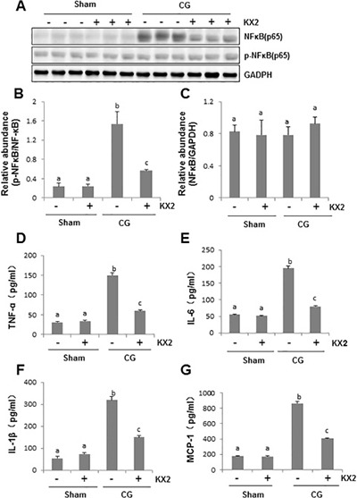
Peritoneal membrane was collected at 21 days after CG injury with or without administration of KX2-391(KX2) (A–G). (A) Peritoneum tissue lysates were subjected to immunoblot analysis with specific antibodies against p-NF-κB (p65), NF-κB(p65) or NADPH. (B) Expression level of p-NFκB(p65) was quantified by densitometry and normalized with NFκB(p65). (C) Expression levels of NFκB(p65)were quantified by densitometry and normalized with GAPDH. Graphs show the expression level of TNF-α (D), IL-6 (E), IL-1β (F), and MCP-1 (G) by ELISA. Data are means ± S.E.M. (n = 6). Bars with different letters (a–c) are significantly different from one another (P < 0.05).