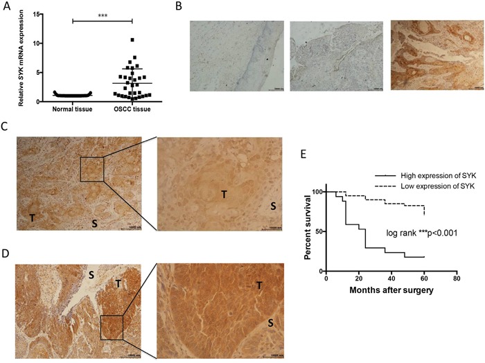
Figure 1. SYK is significantly over-expressed in cancerous tissues and assocaited with overall survival.

(A): Relative expression of SYK in OSCC tissues in comparison with adjacent, histologically normal tissues (n=31). The SYK mRNA level in tumor tissues was normalized to normal tissues. (B): Staining of SYK in normal tissues (left). Staining of SYK in OSCC tissues, SYK-negative (middle) and SYK-positive (right) expression in epithelial layers. (magnification, ×100). (C and D): Different patterns of SYK expression (C): Cytoplasmic and nuclear staining; (D): Predominantly cytoplasmic staining; T, tumor; S, stroma; magnification, ×100 left and ×400 right) in OSCC tissues. (E): Kaplan-Meier curves for the overall survival rate of p OSCC patients were calculated based on SYK expression (high versus low) determined by IHC. Log-rank test was used to compare the significance of the two curves.
