
Figure 3. Piceatannol attenuates proliferation, migration, and invasion of OSCC cell line.
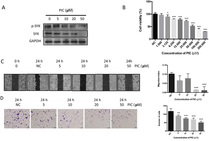
(A): After induced by PIC and vehicle for 24 h, CAL27 cell lysates were harvested and performed Western blot for levels of phosphorylated, total SYK, and GAPDH. (B): CAL27 cells, seeded in 96-well plate for overnight, were stimulated by gradient concentration of PIC (from 1.56 to 200 μM) and the vehicle. Cell viability was detected by CCK8 assay. (C): Representative images (left) were obtained from the wound healing assay induced by PIC. Quantitative analysis of the migration was indicated using migration index (right). (D): Representative images (left) and the quantitative analysis (right) showed the invasion ability of CAL27 cells after induced by PIC using transwell invasion assay. ** p < 0.01; *** p < 0.001.