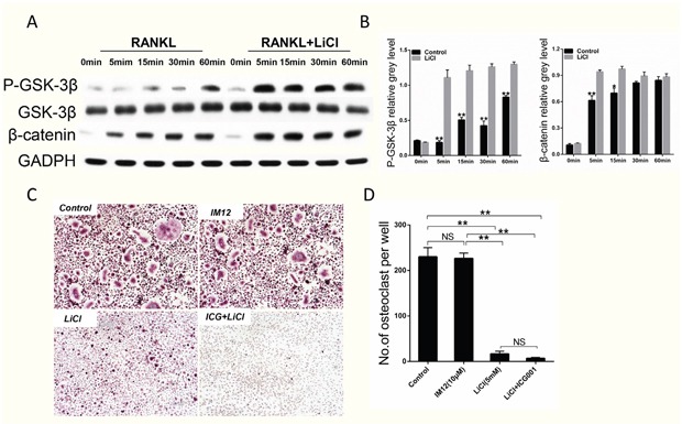
Figure 6. LiCl activation of the GSK-3β/β-catenin pathway did not inhibit RANKL-induced osteoclastogenesis.

(A) After pretreatment with 5 mM LiCl for 4h, RAW264.7 cells were stimulated by 50 ng/ml RANKL for the indicated times (0, 5, 15, 30, 60 min). The cells were then collected and lysed for western blot assay. (B) The relative greys corresponding to Ser9-GSK-3β phosphorylation and β-catenin were quantitated by Image J 6.0 software. (C) After pretreatment with IM12 or LiCl in the presence or absence of ICG-001, BMMs were cultured in induction medium for 5 days. Subsequently, the cells were fixed and stained for TRAP. The control group was not treated. The pictures were taken using a high-quality light microscope at a magnification of 10×. (D) TRAP-positive cells with nuclei ≥ 3 were quantified. (NS: P>0.05; *P<0.05; **P<0.01).
