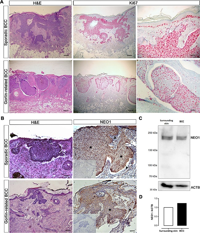
Figure 3. NEO1 is expressed in human BCC.

(A) H&E of sporadic and Gorlin syndrome-related BCC biopsies (bar = 1000 μm). IHC of Ki67 (pink stain) shows highly proliferative tumor cells (bar = 1000 μm and 250 μm). Images are representative photographs of n = 32. (B) H&E of sporadic and Gorlin syndrome-related BCC biopsies (bar = 1000 μm). IHC analysis of NEO1 shows expression (brown staining) in nodules of human BCC biopsies both in the bulk of tumors (asterisk) and palisade (arrow). Hematoxylin (blue) counterstain was used to distinguish nuclei. Negative control of IHC is shown as an insert. Images are representative photographs from n = 32 for sporadic BCC and n = 4 for Gorlin syndrome-related BCC (bar = 500 μm). (C) NEO1 expression in a non-aggressive sporadic human BCC and its healthy surrounding skin was evaluated by WB, ACTB is shown as a loading control. (D) Graph depicting the levels of NEO1 in BCC compared to its surrounding skin evaluated in (c) and normalized by ACTB expression.
