
Figure 5. GPER- and oxygen-dependent regulation of VEGF expression by estrogen.
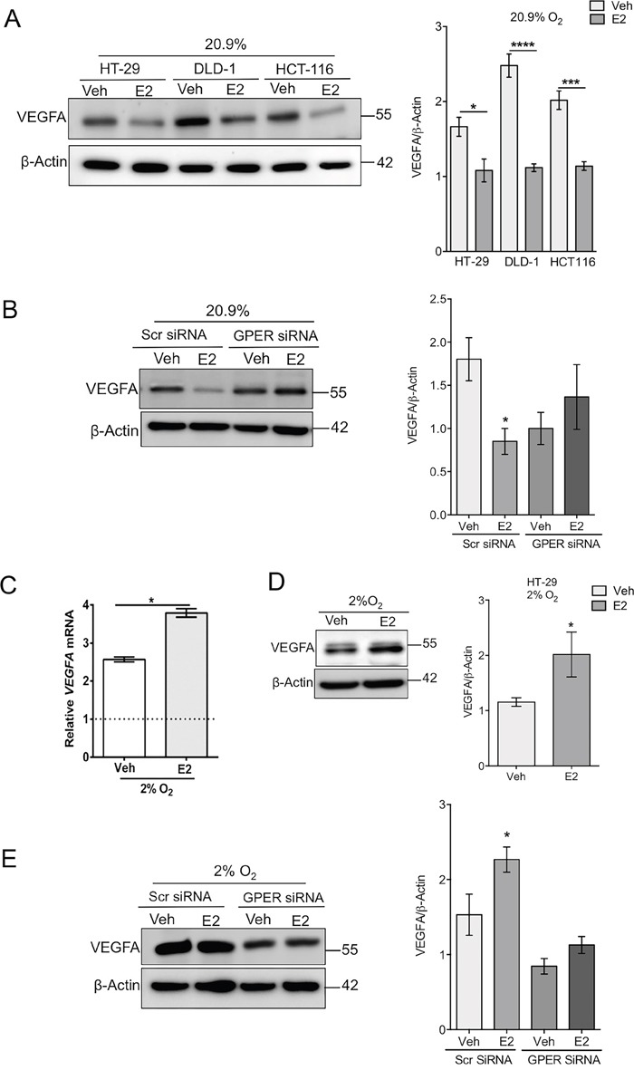
(A) Western blot and densitometry analysis of VEGFA protein expression in a panel of cell lines (HT-29, DLD-1 and HCT116) treated with ethanol control (Veh) or 10nM estradiol (E2) under normoxic conditions. β-actin was used as a loading control. Mean ± SEM, n=4, *P<0.01, ***P<0.001, ****P<0.0001, p-values relative to vehicle treated controls. (B) Western blot and densitometry of VEGFA protein expression in normoxic transfected HT-29 cells treated with vehicle or estradiol. Scrambled control (Scr) or GPER-targeting siRNA were used. Mean ± SEM, n=4, *P<0.05, p-value relative to vehicle treated control. (C) VEGF mRNA expression normalised to PPIB endogenous control in HT-29 cells treated with ethanol (Vehicle) or 10nM estradiol (E2) under hypoxic conditions (2% O2). Dotted line represents vehicle-treated normoxic HT-29 cells. Mean ± SEM, n=3, *P<0.01, p-value relative to vehicle treated control. (D) Western blot and densitometry analysis of VEGF protein expression in hypoxic HT-29 cells treated with ethanol (Veh) or 10nM estradiol (E2). β-actin was used as a loading control, mean ± SEM, n=5 *P<0.05. p-value relative to vehicle treated control. (E) Western blot and densitometry of VEGFA protein expression in hypoxic transfected HT-29 cells treated with vehicle or estradiol. Scrambled control (Scr) or GPER-targeting siRNA were used. Mean ± SEM, n=5 *P<0.05. P-value relative to vehicle Scr treated control.