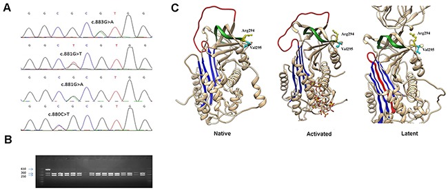
Figure 1. Identification of a mutational hotspot in SERPINC1.

(A) Electropherogram of samples carrying all mutations affecting antithrombin Arg294 and Val295 residues. (B) Restriction fragments generated with BstUI on the PCR product of SERPINC1 exon 5 using the primers described in Materials and Methods. (C) Localization in the structure of antithrombin of affected residues in the native, activated, and latent configurations. The central A sheet is colored in blue, the reactive center loop in red, and s1B in green. Model building was performed by using SWISS-MODEL and the Swiss-PdbViewer programs [Guex N, Peitsch MC. SWISS-MODEL and the Swiss-PdbViewer: an environment for comparative protein modelling. Electrophoresis 1997; 18: 2714–23] (http://www.expasy.ch/spdbv).
