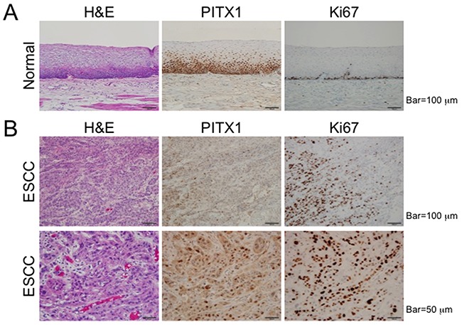
Figure 2. Decreased expression of PITX1 in ESCC.

Representative images produced from formalin-fixed, paraffin-embedded samples of normal mucosa (A) and ESCC (B) stained with H&E, anti-PITX1, or anti-Ki67 antibodies.

Representative images produced from formalin-fixed, paraffin-embedded samples of normal mucosa (A) and ESCC (B) stained with H&E, anti-PITX1, or anti-Ki67 antibodies.