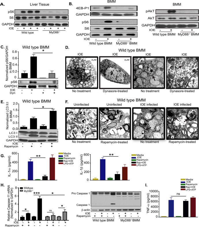
Fig 4. MyD88 inhibits autophagy induction and activates inflammasome via activation of mTORC1 pathway in macrophages.
(A) Representative immunoblots of phosphorylation of S6 (pS6) and total S6 (S6) in whole liver lysates from uninfected and IOE-infected WT or MyD88-/- mice on day 7 p.i. (B) Representative immunoblots showing expression of 4E-BP1, phospho- S6 (pS6), total S6 (S6), phospho-AKT (pAKT), and total AKT in uninfected and IOE-infected WT and MyD88-/- BMM. (C) Representative Immunoblots showing phosphorylation of S6 (pS6) in WT-BMM infected with IOE and treated with/without Dynasore (Dyn). pS6 normalized to GAPDH at 24h p.i. (D) Transmission electron microscopy images of WT-BMM infected with IOE and treated with/without Dynasore at 24h p.i. Left two panels shows low magnification (scale bar 2μm), and right two panels show high magnification (scale bar 500nM) magnification, respectively. Arrow in no Dynasore-treated cells indicating Ehrlichia morula. Arrow on Dynasore-treated cell indicates normal mitochondrial morphology. (E) Immunoblotting analysis showing the expression of LC3I and LC3II in untreated and rapamycin-treated, uninfected and IOE infected WT-BMM at 24h p.i. The density of LC3II:LC3I bands were quantified and normalized to GAPDH loading control, and the ratio of normalized LC3II:I band is shown. (F) Transmission electron microscopy of uninfected (left two panels) and IOE-infected (right two panels) WT-BMM at 24h p.i., in the presence or absence of rapamycin showing higher formation of multiple double membrane autophagosomes in both uninfected and infected cells upon rapamycin treatment (Arrows). All panels are of high magnification (scale bar 500nM) (G) Concentrations of IL-1α and IL-1β in culture supernatant from uninfected and IOE-infected WT-BMM in the presence/absence of rapamycin at 24h p.i. LPS + ATP used as positive control. (H) mRNA expression of Caspase-11 and immunoblot of pro- and active/cleaved caspase-1 in uninfected and IOE-infected WT and MyD88-/- BMMs in the presence/absence of rapamycin at 24h p.i. (I) TNF-α in culture supernatant from uninfected and IOE-infected WT-BMM infected with/without rapamycin treatment at 24h p.i. Negative controls included uninfected cells with or without rapamycin, while positive control included WT-BMM stimulated with LPS and ATP that activates NLRP3 inflammasome. All results are presented as mean ± SD (* P<0.05, **P<0.01, ***P<0.001) from three independent in vitro experiments. ns = not significant. Quantification of immunoblot bands is presented as mean pixel density of bands/GAPDH from three mice/group and is representative of three independent experiments.

