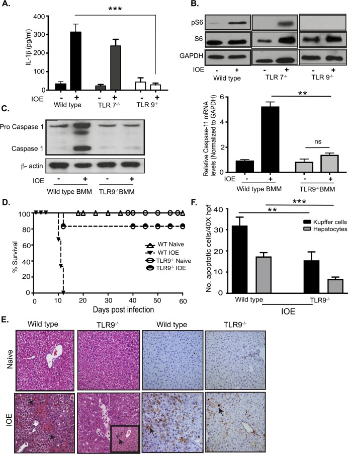
Fig 6. TLR9-dependent inflammasome and mTORC1 activation in IOE infected macrophages.
A) IL-1β in cell culture supernatants from uninfected and IOE infected (MOI 5) WT, TLR7-/-, and TLR9-/- BMM at 24h p.i. (B) Representative Immunoblot analyzing phosphorylation of phospho S6 (pS6) in WT, TLR7-/-, and TLR9-/- BMM at 24h p.i. GAPDH used as loading control. (C) Immunoblot analysis of Caspase-1 in WT and TLR9-/- BMM at 24h p.i. β-actin used as loading control. mRNA expression of Caspase-11 in infected WT and TLR9-/- BMM and controls at 24h p.i. (D) Survival of uninfected and IOE-infected WT and TLR9-/- mice. Data showing 85% survival of IOE infected TLR9-/- mice compared to WT mice till day 60 p.i. (n = 6/group). (E) Representative H&E and TUNEL staining of liver sections from naïve/uninfected and IOE-infected WT and TLR9-/- mice on day 7 p.i. The insert in the H&E staining of liver section from IOE-infected TLR9-/- mice demonstrates an enhanced cellular infiltration consistent with regenerative changes. (F) Quantification of TUNEL positive Kupffer cells and hepatocytes per high power field (HPF) in TLR9-/- and WT mice. Data from in vitro experiments are representative of three independent experiments. All results are presented as mean ± SD (* P<0.05, ***P<0.001).

