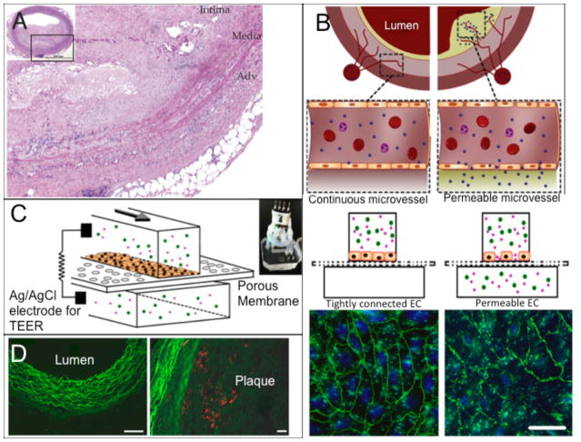
Figure 3.
(A) In vivo study exploring the effect of atherosclerotic lesions on microvessels surrounding an affected artery and on thrombotic complications from microvessel occlusion found dysfunctional endothelium in the surrounding microvessels. (B) Schematic of surrounding microvessels being permeable to nanoparticles due to disrupted endothelium. Fluorescent image shows the normal (left) and disrupted (right) adherens junctions. (C) A schematic of the microfluidic system studying nanoparticle translocation. (D) In vivo results from atherosclerotic aorta from a rabbit being treated with nanoparticles seen in red. Left is the lumen area and right shows the plaque with accumulation of nanoparticles. Reproduced from Sluimer et al. 64 and Kim et al.21 with permission.

