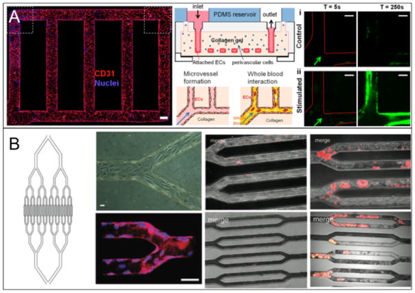
Figure 4.
(A) A microvasculature system that shows occlusion due to aggregated platelets labeled with green CD41a. The endothelialized system where CD31 is shown in red and nuclei are shown in blue (left), a schematic of the entire system (middle) and the occlusion results for both stimulated and non-stimulated vasculature (right) are given. (B) A microvasculature-on-a-chip system that also shows occlusion in thrombotic environments. This study features HLMVECs and whole blood dyed with R6G that preferentially stains platelets and leukocytes. A schematic with endothelialized channels (left half) and platelet occlusion results (right half) are given. The results show occlusion progression with the effects of TNF-α activation for HLMVECs (top) and STX-2 activation for HUVECs (bottom). Reproduced from Zheng et al.24 and Tsai et al.23 with permission.

