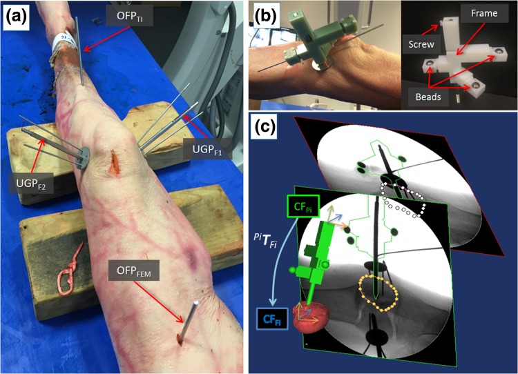
Figure 6.
Intra-operative navigation. Cadaveric specimen with orthopedic pins inserted (a); example of a registration tool RTUGP inserted into UGP pin (b); 2D/3D registration framework (c): 6DOF pose of the fluoroscopic images is estimated using the CAD model of the registration tool and the pin (green object); CT-generated model of the bone fragment (red object) is then registered with the fluoroscopic images; the relative pose between the coordinate frames of the fragment (CFFi) and the inserted pin (CFPi) is defined by the homogeneous transformation Pi T Fi.

