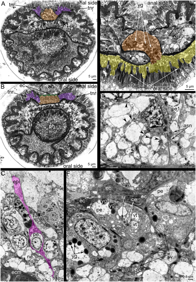
Figure 5.
Organization of the dorsal ganglion and tentacular nerve ring in Phoronis ovalis. Semithin (A,B,D) and ultrathin (C,E,F) transversal sections. (A) Section of the tip of the dorsal ganglion (brown), (B) section of the dorsal ganglion (brown) base: the layer of envelope cells (ec) is marked by green, (C) at the tip of dorsal ganglion, perikarya are surrounded by thin projections of envelope cells (pink), (D) the connection between the dorsal ganglion (brown) and inner ganglion (yellow); (E) a portion of the tentacular nerve ring: perikarya of second type form thin projections (arrowheads), which form bundles (asn), (F) organization of perikarya of middle zone of the dorsal ganglion: perikarya contain yolk granules (yg), centrioles (ce), and synaptic vesicles (sv). Abbreviations: ah, anal hill; ce, centriole; dg, dorsal ganglion; ec, envelope cell, ecm, extracellular matrix; epi, epithelium; gc, gland cells; ig, inner ganglion; m, mouth; mt, mitochondria; ne, neurite; pe, perikaryon; pe1, perikaryon of first type; pe2, perikaryon of second type; ph, pharynx; sv, synaptic vesicle; tnr, tentacular nerve ring; vt, multivesicular body; yg, yolk granule.

