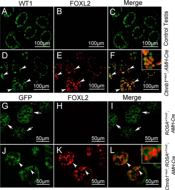
Figure 2.

FOXL2 was expressed in Ctnnb1-overactivated Sertoli cells. WT1 and FOXL2 double staining experiment was performed on E17.5 control and Ctnnb1+/flox(ex3) AMH-Cre testes. WT1 protein (green) was expressed in Sertoli cells of control (A and C) and Ctnnb1+/flox(ex3) AMH-Cre (D and F) testes. FOXL2 protein was detected in remnant seminiferous tubules of Ctnnb1+/flox(ex3) AMH-Cre mice (E, white arrowheads), but not in control testes (B). FOXL2 and WT1 were co-localized in Ctnnb1+/flox(ex3) AMH-Cre testes (F, inset). GFP signal was detected in the Sertoli cells of both control (ROSAmT/mG AMH-Cre, G and I, white arrows) and ROSAmT/mG Ctnnb1+/flox(ex3) AMH-Cre mice (J and L, white arrows). FOXL2 protein (14) was only expressed in ROSAmT/mG Ctnnb1+/flox(ex3) AMH-Cre mice (K, white arrowheads), but not in ROSAmT/mG AMH-Cre testes (H), and co-localized with GFP protein (L, inset).
